WE WILL LEARN ABOUT LENSES, AND EYE, MIRRORS ALSO!!
TERMS RELATED TO SPHERICAL MIRRORS

In a Concave mirror parallel rays after reflection meet at a point on the Principal Axis which is known as Focus/Focal Point. It is denoted by letter F.
Meanwhile, in a Convex mirror parallel rays after reflection appear to meet at a point which is know as Focus/Focal Point. It is denoted by letter F.
The centre of a reflecting surface of a spherical mirror is called it’s Pole. It is denoted by the letter P.
The centre of the hollow sphere of which the spherical mirror is a part of is called it’s Centre of Curvature. It is denoted by the letter C.
The radius of the hollow sphere of which the spherical mirror is a part of is called it’s Radius of Curvature. It is denoted by the letter R.
The straight line passing through the centre of curvature (C) and the pole (P) of the spherical mirror is called the Principal Axis. Denoted by PA.
The Normal at any point of the spherical mirror is the straight line drawn perpendicularly to the centre of curvature (C) of the mirror. Denoted by letter N.
The distance between the pole and the focus is called the spherical mirror’s Focal Length.
Focal length = 1/2*Radius of Curvature
f = R/2 where R denotes the Radius of Curvature and f denotes the Focal Length
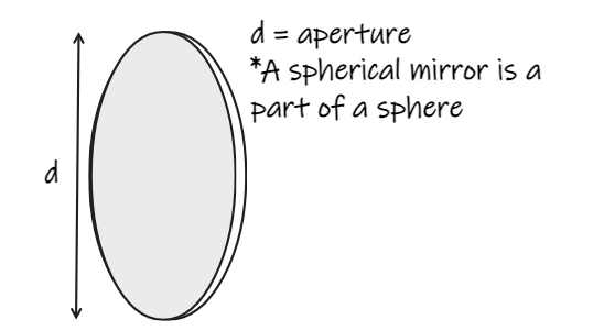
The width of a spherical mirror from which reflection can take place is called it’s Aperture.

An imaginary plane passing through the focus and at right angles to the principal axis is called focal plane

When the rays of light after getting reflected from a mirror (or after getting refracted from a lens) actually meet at a point, a real image is formed which can be obtained on a screen.
When the rays of light after getting reflected from a mirror (or after getting refracted from a lens) APPEAR to meet at a point, a virtual image is formed. Such an image can only be seen through a mirror/ a lens but can’t be obtained on a screen.
CONVEX MIRROR

This is a curved mirror which always forms a virtual, erect and diminished image. This mirror diverges light rays so they never meet, therefore, since light rays never meet, the image is virtual. It provides an erect image. The reflected ray appears to have come from the focus of the mirror (point of convergence of light rays). If the position of an object is at infinity (meaning; very far; more than 20 feet or more), the nature of the image formed will be virtual and erect. The size of the image will be VERY small (point sized). If the position of an object is between pole and infinity, the image formed will, was always, be virtual, erect and diminished, but the size of the image will be smaller than the object, but NOT TOO MUCH SMALLER than the object.
Uses of Convex mirror:-
1. Since it has a very wide field of view, they are used as rear view mirrors and side view mirrors in vehicles.
2. It is also used as traffic mirrors along roads and in parking lots due to the very wide field of view.
CONCAVE MIRROR

This is also a curved mirror which forms many types of images.
Following are the cases of object placement and the type of image formed:-

DISPERSION
It is a phenomenon through which white light splits into its constituent colours VIBGYOR, which are :-
- Violet
- Indigo
- Blue
- Green
- Yellow
- Orange
- Red
When a white light is passing through a prism, violet bends the most while red bends the least, explaining why we can see different colours once we disperse white light which are heterogeneous in nature (do not completely mix with each other; are separate but still are a mixture)
In other word, dispersion occurs when white light is incident on a prism. Rainbow is a natural phenomenon which is caused due to dispersion.
V = f*λ
Here, V is the speed of light, f is the frequency and λ is the wavelength in meters.
LENSES

A lens is a portion of a transparent refracting medium bounded by a pair of surfaces.
TYPES OF LENSES

There are two types of lenses :-
- Convex or Converging lens
- Concave or Diverging lens
There are three types of Convex lenses :-
- Double Convex (Or just Convex)
- Plano Convex
- Converging Convex
There are also three types of Concave lenses :-
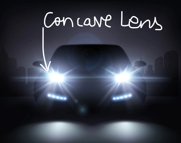
- Double Concave (Or just Concave)
- Plano Concave
- Diverging Concave
Terms regarding lenses :-
- Centres of Curvature – Each side of a lens can be referred to as a part of a sphere. The centres of curvature will be the centre of curvature of each sphere.
- Radii of Curvature – Each radius of each sphere are known as the Radii of Curvature.
- Principal Axis – When we draw a straight line joining the centres of curvature of both the spheres, we call it as the Principal Axis of the lens.
- Optical centre – The optical centre of the lens is defined as the centre of the intersecting point of both the spheres.
- Principal Focus/Focal length – When a beam of parallel rays (parallel to the principal axis) falls on the lens, they either all converge at a point on the principal axis, or appear to diverge from a point on the principal axis (second principal focus). The focal length is equal to the distance of the second principal focus from the optical centre of the lens.
RULES FOR IMAGE FORMATION IN LENSES

- An incident ray which is parallel to the principal axis passes through (or appears to come from) the second principal focus of the lens.
- An incident ray passing through the optical centre of the lens goes undeviated
- An incident ray passing through the principal focus of the lens are appearing to pass through it will become parallel to it after refraction from the lens.
IMAGE FORMATION BY CONVEX LENSES
- If an object is very far off, a convex lens will form a real, diminished and point sized image at the focus of the lens.
- If an object is placed beyond the centre of curvature , the image will be formed between the focus and twice the focus only, and will be real, inverted and diminished
- If an object is place at the centre of curvature of the lens, it is formed at the point defined by twice the focus only, and will be real, inverted but of the same size as the object.
- If an object is placed between the centre of curvature and the focus of the lens on the other side, it will form a real, inverted and enlarged image beyond the 2F (length equal to the radii of curvature on the other side) point of the lens
- If an object is kept at the first Principal focus of the lens, it will form a real, inverted and magnified image formed very far off or in other words, at infinity.
- If an object is kept between the optical centre and the focus of the lens then the image will be formed in front of the lens, it will be virtual and erect, along with being enlarged.
IMAGE FORMATION BY CONCAVE LENSES
A concave lens always forms a virtual, erect and diminished image which appears to be located between the Optical centre and the Focus of the lens on the same side as the object is.
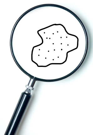
FUN FACT: A convex lens is also used in a magnifying glass or a microscope for magnification too !!!
EYEEEEEE!!!!!!!!!!!!!!!!


The human eye helps us see and has many parts which have a specific function.
- Sclerotic- It is the outermost covering of the eye ball. It is made up of ”tough tissues” which are white in colour. It helps to house and protect the internal organs of the eye.
- Cornea- It is the front bulging part of the eye and is made up of transparent tissues. This is the part which allows light to enter. Most of the refraction of light occurs at the Cornea itself.
- Choroid- It is a grey membrane attached to the sclerotic from the inner side. It darkens the eye from inside so that internal reflection cannot occur. If it occurs, then our brain will not be able to perceive the image.
- Optic Nerve- It is a bundle of approximately 70,000 nerves which help in carrying optical messages to the brain in the form of electrical signals. They originate from the brain and enter into the eye ball from behind.
- Retina- It is the photosensitive layer at the back of the eye and is the site of image formation. It contains two types of cells- Rods which control scotopic vision (helping to sense dim light) and Cones which control photopic vision (helping to sense colour, or, bright light). It receives the optical image of the object and then converts it to optical pulses (electrical signals). The optical image of the object is real and inverted in nature.
- Yellow spot- It is a small area facing the crystalline lens and has a lot of nerve endings. It is also slightly raised and yellow in colour. It helps in sending a large number of optical pulses to the brain allowing the brain to process a more clear and concise view in the form of electrical signals.
- Blind spot- It is a region on the retina from where the optic nerve enters the eye ball. It does not have a nerve ending and is insensitive to light.
- Crystalline lens- It is a double convex lens made up of transparent tissues. It helps in focusing the images clearly on the retina by providing a finer adjustment of focal length
- Ciliary muscles- They hold together the Crystalline lens in it’s place. They also help in increasing and decreasing the focal length of the crystalline lens so that the images of the objects placed at different angles are also clearly focused on the retina.
- Iris- It is a circular diaphragm in front on the crystalline lens and imparts the so called “colour of the eye” to the eye due to the heavy amounts of pigmentation. It has tiny muscles which are arranged around the pupil in a circular manner which helps to increase or decrease the diameter of the same. It controls the amount of light entering the eye by decreasing or increasing the diameter of the pupil.
- Pupil- It is a small hole in the eye’s centre which regulates and controls the amount of light entering into the eye.
- Vitreous Humour- It is a dense, jelly-like fluid, which is slightly grey in colour and fills the part of the eye between the crystalline lens and the retina. It prevents the eye ball from collapsing due to the change in atmospheric pressure and also helps in focusing light rays clearly on the retina. It is named so because it is water insoluble.
- Aqueous Humour- It is a watery saline fluid, which fills the part of the eye between the crystalline lens and the cornea. It prevents the front part of the eye from collapsing due to the change in atmospheric pressure and helps keep the cornea moist.
Now, you may also think that how can an eye collapse?
Well, eyes are jelly-like structures, so they can easily collapse due to the change in atmospheric pressure. Change in atmospheric pressure can cause an increase in blood pressure which can result in the collapsing of the eye.
This is prevented by Aqeuous humour.
Range of vision

Far point is defined as the maximum range through which a human can see; it is infinity for a normal human eye.
Near point is defined as the nearest distance from where the eye can focus clearly; it is 25 cm for a normal human eye.
Range of vision is defined as the distance between the far and near point
The power of accommodation of the eye is defined as the ability of the eye lens to adjust it’s focal length, which cannot be decreased beyond a certain limit, which, in this case, lies when it wants to focus on an object that is less than 25 cm away from the eye.
DEFECTS OF VISION

- Myopia- Far off vision of a person is blurred in this condition since the image is formed in front of the retina. Thus this indicates that we need to diverge the light ray more. We can use a concave/Diverging lens for that.
- Hypermetropia- Nearby vision of a person is blurred since the image is formed behind the retina. Thus this indicates that we need to make the light rays converge early. We can use a convex/Converging lens for that.
- Presbyopia- It is a combination of myopia and hypermetropia and occurs in people who are in the old ages of their life span, due to the weakening of ciliary muscles. A bifocal lens consisting of a convex and a concave lens can be used.
CATARACT

The fluid in the eye solidifies with age. This causes for the eye lens to become milky and cloudy. This condition is known as Cataract and can be corrected using surgery. If left untreated, it may result in partial/complete vision loss.
BRAILLE SYSTEM
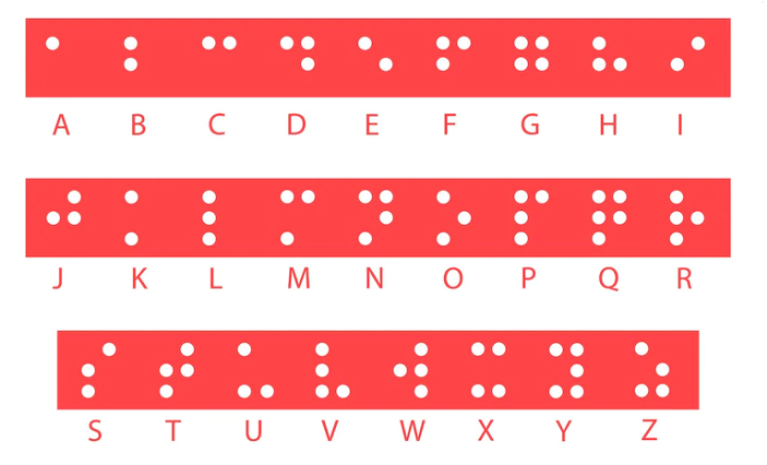
This was developed in 1824 by Louis Braille (a blind person) and was adopted in 1932
It consists of a code of 63 characters and are arranged in a six-position matrix or cell.
They are embossed on Braille Sheets and are read by the visually challenged through the sense of touch.
The dots are raised slightly.
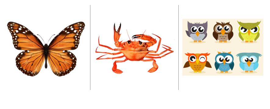
ANIMALIC VISION!!!
Vision in some animals can be described in the following ways:-
- Crab – It has 2 small eyes placed in such a way that it can see all around
- Butterfly- It has compound eyes. Which means that each eye is made up of thousands of smaller ones. It’s eyes are placed in such a way that it can see all around it’s surrounding.
- Owl- It has a large cornea and a large pupil to allow more light to enter it’s eyes. It has more rods than cones.